Student Research
Comparing Mitosis and Meiosis
How cellular context allows the same proteins to act dramatically differently
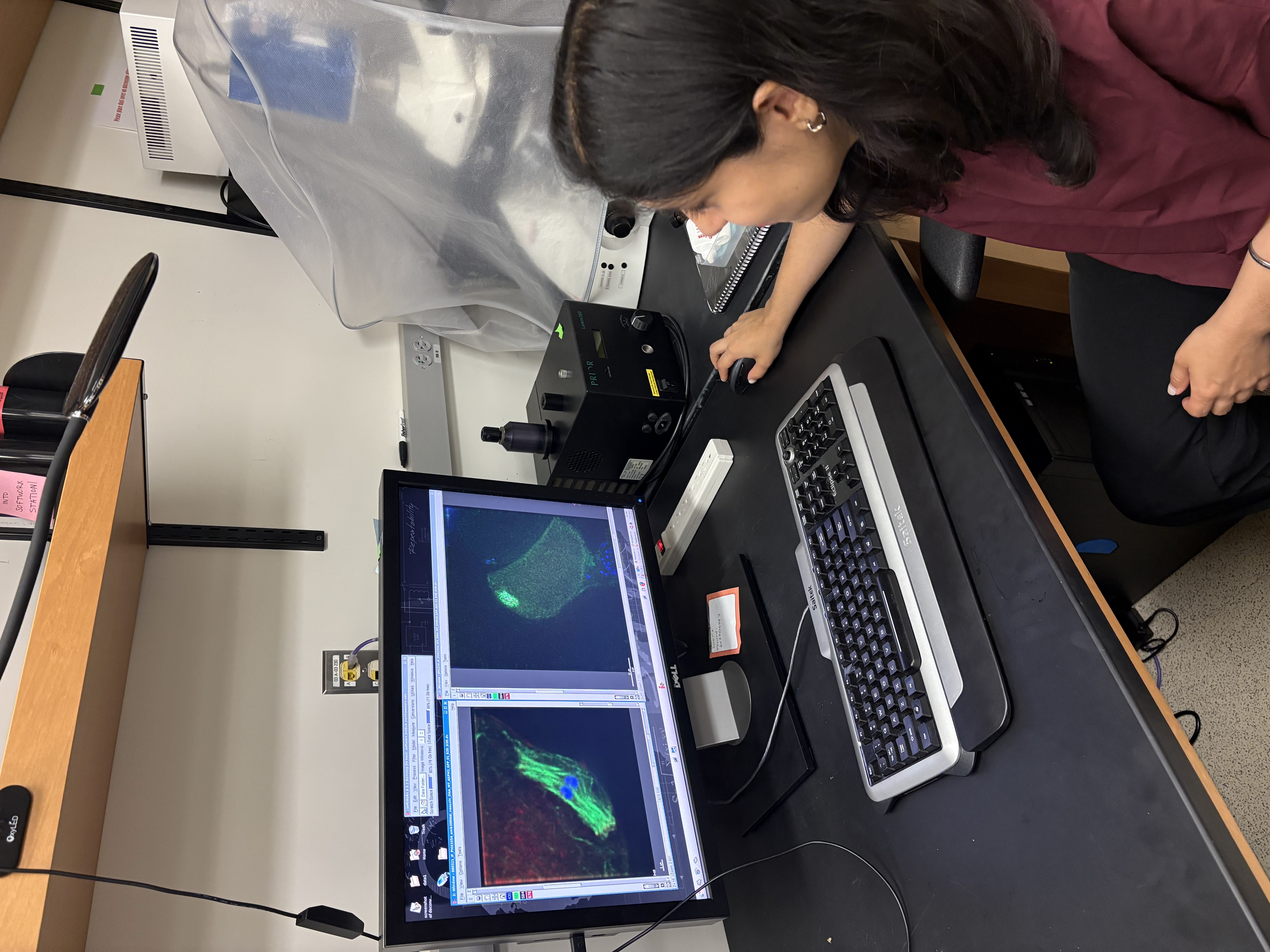
In most cell types, centrosomes mediate cell division by providing organizational cues for the formation of a microtubule-based bipolar spindle. Centrosomes are comprised of a centriole pair and surrounding pericentriolar material (PCM) that helps to both assemble the spindle by nucleating microtubules and maintain bipolarity by organizing the microtubule minus ends into two poles. In contrast, oocyte meiosis is a specialized form of cell division in which centrosomes are typically absent. Nonetheless, bipolar spindles still form, demonstrating that alternate mechanisms must be utilized in oocytes to ensure faithful meiosis.
Our research utilizes Caenorhabditis elegans as a model to investigate these mechanisms. Polo-like kinases are critical regulators of mitotic chromosome segregation and spindle formation across species. In C. elegans, polo-like kinase 1 (PLK-1) promotes centrosome maturation during mitosis and is required for nuclear envelope breakdown in oocytes. How PLK-1 contributes to spindle assembly and whether it also promotes spindle stability during oocyte meiosis were not known. To address these questions, we utilized rapid auxin-inducible degradation to deplete PLK-1 specifically from oocytes, and found that PLK-1 is required for spindle assembly and spindle stability. Interestingly, these experiments also revealed novel roles for this conserved kinase in oocyte meiosis that are distinct from its functions in mitosis. Specifically, we found that PLK-1 depletion resulted in excess microtubule nucleation throughout the oocyte, and premature PCM recruitment to the sperm-provided centrioles. During mitosis, one of PLK-1’s major functions is to recruit PCM, rather than block PCM accumulation. Our discovery is therefore paradigm-shifting, since it shows that PLK-1 can either promote or suppress centrosome maturation, depending on the context.
Altogether, these findings identify PLK-1 as a key player in acentrosomal oocytes that promotes the faithful execution of the meiotic divisions. Furthermore, this work elucidates a poorly understood form of cell division that is essential for sexual reproduction, allowing for genetically diverse progeny. Errors in oocyte meiosis increase with maternal age; therefore, understanding the mechanisms underlying faithful meiosis can indicate potential solutions for infertility. To read more, see Narula and Wignall (2025) – PLK-1 suppresses centrosome maturation and microtubule polymerization to ensure faithful oocyte meiosis. – Juhi Narula
Hot on the Trail of Evolution
Revealing how neurobiological changes drive the evolution of temperature preference behavior in flies
 From harsh deserts to frozen mountaintops, animals have found a way to thrive in the many climatic niches found on our planet. As species adapt to new thermal environments, they evolve molecular and physiological mechanisms for survival, but also changes in temperature and humidity preference that reflect the conditions of their new habitat. However, we know little about how animal behavior evolves, and how differences in thermosensation underlie these behavioral adaptations. Insects, small cold-blooded animals which comprise the vast majority of species biodiversity, offer ideal models for the study of evolutionary responses to environmental factors.
Insects rely on their thermosensory system to sense and respond to external temperature: an array of specialized sensory neurons equipped with molecular heat receptors that detect changes in external temperature and send signals to the brain, where this information is then processed to guide behavior.
In my research, we discovered that at least two distinct neurobiological mechanisms drive the evolution of temperature preference behavior in flies of the genus Drosophila. Fly species from mild climates (D. melanogaster and D. persimilis) avoid heat, and we show that changes in the sensitivity of their heat receptor adjust avoidance behavior to match the conditions of their natural habitats. By contrast, the desert-adapted fly D. mojavensis is actively attracted to heat, mediated by the same heat receptor. Instead, this attraction behavior is driven by specific changes in how heat signals are processed in the brain.
My work illustrates how changes in both sensory neurons and brain circuits can contribute to the insect's ability to adapt to different thermal habitats. Ultimately, my hope is that my findings may help us better understand and perhaps predict how climate change will affect the distribution of insects and the many ecological systems that depend on them. For more details, see Capek, et al., Evolution of temperature preference in flies of the genus Drosophila. – Matthew Capek
Key Players of Gene Positioning
Uncovering the noncanonical role of a nuclear export protein in gene targeting
 The nuclear pore complex (NPC) interacts with the genome to regulate genome architecture and integrity as well as gene expression. The NPC is composed of ~30 different nuclear pore proteins or nucleoporins (Nups), which physically associate with hundreds to thousands of sites in the genome to modulate transcription in budding yeast, flies, and mammals. In yeast, these Nups can target activated inducible genes to the nuclear periphery, where they experience an increase in transcription. This peripheral localization is not dependent on transcription; however, it requires various players such as transcription factors (TFs), TF-binding sites known as DNA zip codes, Nups like Nup2, and—from our recent findings—exportin-1 (XPO1 or Crm1 in yeast). Using a previously found mutation in a TF that blocks peripheral targeting of various genes, we performed quantitative proteomics and found that the mutation reduced interactions between the TF and Crm1/XPO1. Crm1 is a large essential nuclear export receptor whose known function is shuttling large cargo proteins and RNAs out of the nucleus and into the cytoplasm. When probing the localization of Crm1 in vivo, we not only found a high correlation between Crm1 and Nup binding sites genome-wide and at the enhancers of highly active genes, but also a requirement for Crm1 to mediate interactions between Nups and the genome. Inhibition or loss of Crm1 or Nup2, respectively, causes a major decrease in nascent transcription globally. My work suggests that Crm1 docks TF-bound enhancers at the NPC to enhance transcription. For more details, see: Exportin-1 functions as an adaptor for transcription factor-mediated docking of chromatin at the nuclear pore complex. – Tiffany Ge
Unlocking the T-box
RNA form meets function
T-box riboswitches are specialized RNA elements that regulate genes involved in amino acid metabolism in Gram-positive bacteria. They do so by sensing the aminoacylation state of tRNAs. Unlike other riboswitches that respond to small molecules, T-boxes are uniquely equipped to recognize and bind structured RNA with high specificity, making them a key model for studying RNA-RNA interactions. Their role in regulating essential metabolic pathways also positions them as promising targets for novel antibiotics.
To deepen our understanding of this regulatory mechanism, we investigated the translational regulation mediated by the Mycobacterium tuberculosis (Mtb) IleS T-box riboswitch. Our study revealed that both translational and transcriptional T-boxes share a two-step tRNA binding model: first, the tRNA anticodon is recognized to form a partially bound state, followed by the stable binding of the tRNA 3'-NCCA end. Interestingly, the Mtb IleS T-box exhibits longer-lived partially bound states and greater conformational flexibility compared to transcriptional T-boxes, suggesting its adaptation for translation regulation. We also identified two key structural elements—a pseudoknot in the Stem IIA/B-linker and a conserved RAG sequence—that play critical roles in enhancing the stability of the tRNA-bound state through allosteric interactions and conformational constraints.
These findings offer new insights into the mechanistic differences between transcription- and translation-regulating T-boxes, advancing our understanding of RNA dynamics and their potential as targets for disrupting bacterial gene expression. For further details, see Campos-Chavez et al., Translational T-box riboswitches bind tRNA by modulating conformational flexibility. – Eduardo Campos-Chavez